45 brain mri with labels
Labels · abidikhairi/brain-mri-segmentation · GitHub In this repository Brain MRI Segmentation Using FCM (Labeling) - Stack Overflow Brain MRI Segmentation Using FCM (Labeling) I am doing Brain MRI segmentation using Fuzzy C-Means, The volume image is n slices, and I apply the FCM for each slice, the output is 4 labels per image (Gray Matter, White Matter, CSF and the background), how I can give the same label (Color) for each material for all the slices) I am using matlab.
Brain MRI: How to read MRI brain scan | Kenhub MRI is the most sensitive imaging method when it comes to examining the structure of the brain and spinal cord. It works by exciting the tissue hydrogen protons, which in turn emit electromagnetic signals back to the MRI machine. The MRI machine detects their intensity and translates it into a gray-scale MRI image.

Brain mri with labels
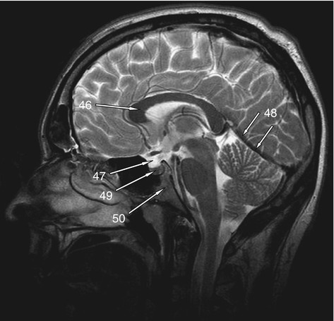
MRI head sagittal T1 - labeling questions | Radiology Case ... The labeled structures are (excluding the correct side): temporal horn of lateral ventricle primary fissure of cerebellum choroid plexus trigone (atrium) of lateral ventricle horizontal fissure of cerebellum occipital horn of lateral ventricle intraorbital segment of optic nerve diploic space of parietal bone body of caudate nucleus maxillary sinus MRI head axial T2 - labeling questions - Radiopaedia The labeled structures are (excluding the correct side): cervical spinal cord posterior arch of C1 odontoid process (peg or dens) of C2 parotid gland intradural segment (V4) of dominant vertebral artery cisterna magna intradural segment (V4) of non-dominant vertebral artery cerebellar tonsil occipital condyle medulla oblongata NITRC: Manually Labeled MRI Brain Scan Database: Tool/Resource Info Manually Labeled MRI Brain Scan Database Visit Website Image 1 of 3 Click for more. This is a continuously growing and improving database of high-quality neuroanatomically labeled MRI brain scans, created not by an algorithm, but by neuroanatomical experts. All results are checked and corrected.
Brain mri with labels. A unified 3D map of microscopic architecture and MRI of the human brain ... 27.04.2022 · The inclusion of five microscopy labels, blockface images, and three quantitative MRI contrasts provides a wealth of anatomical information ().The full-brain coverage allows for detailed and comparative analyses of architectonic features for mapping the cortical laminar structure (20–23).A second important application is the atlasing of small brain structures that … 101 Labeled Brain Images and a Consistent Human Cortical Labeling ... Labeled anatomical subdivisions of the brain enable one to quantify and report brain imaging data within brain regions, which is routinely done for functional, diffusion, and structural magnetic resonance images (f/d/MRI) and positron emission tomography data. Brain charts for the human lifespan | Nature 06.04.2022 · To extend the scope of brain charts beyond the four cerebrum tissue volumes, we generalized the same GAMLSS modelling approach to estimate normative trajectories for additional MRI phenotypes ... Labeled imaging anatomy cases | Radiology Reference Article ... This article lists a series of labeled imaging anatomy cases by body region and modality. Brain CT head: non-contrast axial CT head: non-contrast coronal CT head: non-contrast sagittal CT head: angiogram axial CT head: angiogram coronal CT...
Labeling Brain Structures - John Muschelli 1 Labels in template space. In Processing Within-Visit MRI, we registered the T1 image to the Eve template using a non-linear registration (SyN) (Avants et al. 2008). Also, we applied this transformation to the intensity-normalized T1, T2, and FLAIR images, so that these image are located in the same space as the Eve atlases. We can overlay the ... UCLA Brain Mapping Center - ICBM Template To view both the structural MRI and the labels launch the program typing Display icbm_template.mnc -label icbm_labels_corrected.mnc. The opacity of the labels can be set in the Colour Coding menu. The number of each label appears at the bottom left of the orthogonal views window. MRI Brain Atlas - University of Minnesota This web app Atlas is intended for veterinary students and radiologists seeking quick access to canine brain anatomy through a mobile device. Via a toggle button, either MRI images or approximately comparable Brain Transection images may be viewed with or without labels. Navigation & Labels. Brain Tumor Sequence Registration (BraTS-Reg) Challenge: … Registration of baseline pre-operative (treatment-naïve) and follow-up brain tumor MRI scans is challenging, yet a clinically important task for a multitude of reasons. Brain tissue shows heavy deformations induced by the apparent tumor (also known as mass effect) that following its resection are relaxed due to the relieving pressure from the resected tissue. Such deformations …
Brain Tumor Classification using Machine Learning - DataFlair One such application of deep learning to detect brain tumors from MRI scan images. About Brain Tumor Classification Project . In this machine learning project, we build a classifier to detect the brain tumor (if any) from the MRI scan images. By now it is evident that this is a binary classification problem. Examples of such binary classification problems are Spam or Not spam, … Atlas of BRAIN MRI - W-Radiology Brain magnetic resonance imaging (MRI) is a common medical imaging method that allows clinicians to examine the brain's anatomy (1). It uses a magnetic field and radio waves to produce detailed images of the brain and the brainstem to detect various conditions (2). Brain Tumor MRI Dataset | Kaggle A brain tumor is a collection, or mass, of abnormal cells in your brain. Your skull, which encloses your brain, is very rigid. Any growth inside such a restricted space can cause problems. Brain tumors can be cancerous (malignant) or noncancerous (benign). When benign or malignant tumors grow, they can cause the pressure inside your skull to ... Frontiers | 101 Labeled Brain Images and a Consistent Human Cortical ... Labeled anatomical subdivisions of the brain enable one to quantify and report brain imaging data within brain regions, which is routinely done for functional, diffusion, and structural magnetic resonance images (f/d/MRI) and positron emission tomography data.
MRI scan of brain - Stock Image - P332/0482 - Science Photo Library
CaseStacks.com - MRI Brain Anatomy Labeled scrollable brain MRI covering anatomy with a level of detail appropriate for medical students. Show/Hide Labels. MRI Brain Anatomy. Back to Anatomy Overview. ... Labelled radiographs and CT/MRI series teaching anatomy with a level of detail appropriate for medical students and junior residents. Pelvis. Pelvic MRI anatomy

Dr Balaji Anvekar FRCR: Brain Abscess MRI
Researchers automate brain MRI image labeling, more than ... - ScienceDaily Researchers have automated brain MRI image labeling, needed to teach machine learning image recognition models, by deriving important labels from radiology reports and accurately assigning them to...
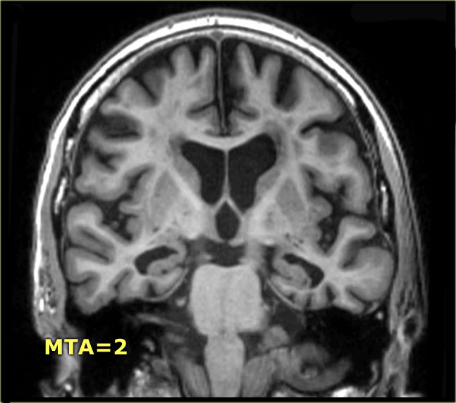
The Radiology Assistant : Brain - Dementia: Role of MRI
Automated MRI image labelling processes 100,000 brain exams in under 30 ... Researchers from the School of Biomedical Engineering & Imaging Sciences at King's College London have automated brain MRI image labeling, needed to teach machine learning image recognition models,...

Dr Balaji Anvekar FRCR: Superior cerebellar territory infarct
Diffusion MRI - Wikipedia Diffusion MRI relies on the mathematics and physical interpretations of the geometric quantities known as tensors.Only a special case of the general mathematical notion is relevant to imaging, which is based on the concept of a symmetric matrix. Diffusion itself is tensorial, but in many cases the objective is not really about trying to study brain diffusion per se, but rather just …

Multiple sclerosis | Image | Radiopaedia.org
Brain lobes - annotated MRI | Radiology Case | Radiopaedia.org brain anatomy by Diana Paduraru; Brain Anatomy by Dr Renor Gonçalves de Castro Neto atlantic fellow session by Ophir Keret; MI 2020-2 by Mariangela Alvarado Molinaro; Brain by Dr Roya Faghani; Ct by Hector De La Cruz; Matrikulasi Koas 17 Januari 2021 by Dr. Gregorius Enrico, Sp.Rad "How To Read a Head CT" Chapter 2 Supplement by Stefan Tigges

Brain MRI Quiz
In vivo brain endocannabinoid metabolism is related to ... - Nature Jul 29, 2022 · In vivo brain endocannabinoid metabolism is related to hippocampus glutamate and structure – a multimodal imaging study with PET, 1 H-MRS, and MRI. Neuropsychopharmacol. (2022). ...
Test 1 | Radiology Key
Anatomical diagrams of the brain - e-Anatomy - IMAIOS 13.09.2021 · A topographical anatomy of the brain showing the different levels (encephalon, diencephalon, mesencephalon, metencephalon, pons and cerebellum, rhombencephalon and prosencephalon) as well as a diagram of the various cerebral lobes (frontal lobe, occipital, parietal, temporal, limbic and insular). Please note that the limbic lobe is functional and thus …

Normal Brain CT MRI Imaging ( In Arabic ) Prof Dr Mamdouh Mahfouz - YouTube
Harvard University Show pointers Show labels Show list All modalities to: ...

MRI BLOG: Pitfalls of Diffusion Weighted Imaging
Brain MRI Dataset | Kaggle Brain MRI Dataset | Kaggle. Haşim Mumcu · Updated 3 years ago. arrow_drop_up. 5. New Notebook. file_download Download (8 MB)

How large is your brain? MRI - YouTube
brain anatomy | MRI coronal brain anatomy | free MRI cross sectional ... ELBOW AXIAL. WRIST AXIAL. WRIST CORONAL. KNEE CORONAL. KNEE SAGITTAL. ARTERIES UPPER LEG. ARTERIES LOWER LEG. This MRI brain coronal cross sectional anatomy tool is absolutely free to use. Use the mouse scroll wheel to move the images up and down alternatively use the tiny arrows (>>) on both side of the image to move the images.

Brain, MRI - Stock Image - C036/6075 - Science Photo Library
MRI Brain Animated Quiz - University of Minnesota MRI Brain Animated Quiz. Canine Brain MRI Anatomy Quiz. Sequentially click/tap: first the dot associated with a term; then, its corresponding target dot on the MRI image. If a line connection appears, your choice was correct! White Matter. Cerebral Cortex. Olfactory Bulb. Longitudinal Fissure.
Psych 104 Chapter 3 Lecture 2
Normal chest MDCT with anatomic labels | e-Anatomy - e-Anatomy … 10.03.2022 · But for educational purposes, we put anatomical labels on the presumed place of these structures. The IASLC lymph node map provides a reproducible and consistent set of definitions for the discussion of regional lymphadenopathy in patients with lung cancer. However, because of its comprehensiveness and text-based presentation, it may be challenging to …
BrainSuite Diffusion Pipeline | BrainSuite
CPT Code for MRI Brain, Breast, Lumbar Spine and Shoulder Find below the latest Radiology CPT codes for for MRI of Brain, Breast, Lumbar Spine and Shoulder: CPT Codes for MRI Lumbar spine In human Lumbar spine is represented by the 5 vertebrae in between the ribcage and the pelvis forming the largest segment of the vertebral column. Depending on the condition that one is treated on these parts of the ...

Brain MRI Interpretation - YouTube
MRI anatomy | free MRI axial brain anatomy - Mrimaster.com This MRI brain cross sectional anatomy tool is absolutely free to use. Use the mouse scroll wheel to move the images up and down alternatively use the tiny arrows (>>) on both side of the image to move the images.

The Radiology Assistant : Brain Anatomy | Brain anatomy, Radiology imaging, Mri brain
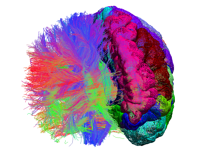
Labeled MRI Brain Scans - Neuromorphometrics We can also label scans that you provide and we are very interested in labeling white matter anatomy as seen in diffusion-weighted MRI scans. If you want an aggregate version of our data, we can provide it as a probabilistic atlas. The cost to label a single scan is $2449 (USD).

Making My Theology Work: An Introduction - ParkingSpace23ParkingSpace23
Brain MRI Atlas on the App Store Brain MRI Atlas is a FREE app that allows you to navigate through hundreds of of labeled brain structures. It is designed for all healthcare professionals as an interactive study and reference tool. Program Features: - Serial sequential axial T2 FLAIR images of the brain. - Structure labels organized by category.

Images of Cadaver and Human Brain of 7.0 T MRI In Vivo | Radiology Key
MRI brain (summary) | Radiology Reference Article - Radiopaedia MRI brain is a specialist investigation that is used for the assessment of a number of neurological conditions. It is the main method to investigate conditions such as multiple sclerosis and headaches, and used to characterize strokes and space-occupying lesions. Reference article
Post a Comment for "45 brain mri with labels"