38 structure of the heart without labels
Heart anatomy: Structure, valves, coronary vessels | Kenhub The heart is shaped as a quadrangular pyramid, and orientated as if the pyramid has fallen onto one of its sides so that its base faces the posterior thoracic wall, and its apex is pointed toward the anterior thoracic wall. diagram of heart without labels 13 Best Images of Hip Anatomy Of The Worksheet - Sunflower Anatomy. 11 Pics about 13 Best Images of Hip Anatomy Of The Worksheet - Sunflower Anatomy : u414adad: heart diagram without labels, Congestive Heart Failure: The Essence of Heart Failure Course | CEUfast and also 13 Best Images of Hip Anatomy Of The Worksheet - Sunflower Anatomy.
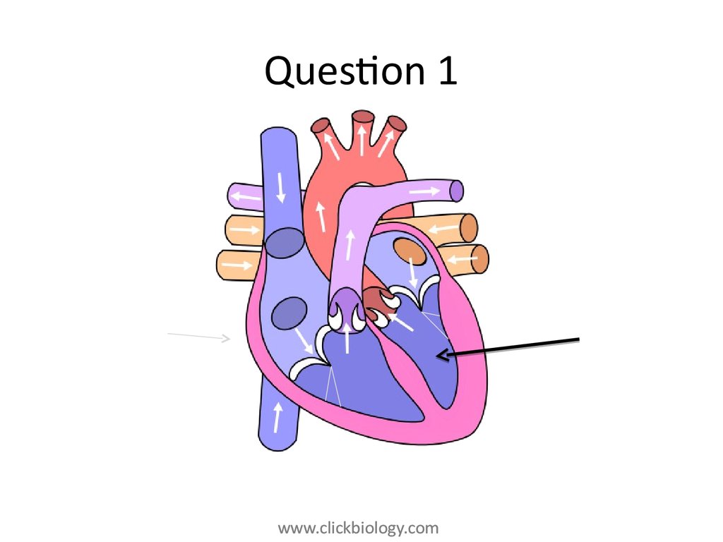
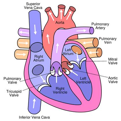
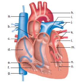
Label the heart — Science Learning Hub In this interactive, you can label parts of the human heart. Drag and drop the text labels onto the boxes next to the diagram. Selecting or hovering over a box will highlight each area in the diagram. Right ventricle Right atrium Left atrium Pulmonary artery Left ventricle Pulmonary vein Semilunar valve Vena cava Aorta Download Exercise Tweet

Structure of the heart without labels
heart without label Vector Winner Golden Label 458912 Vector Art At Vecteezy . winner vector golden label vecteezy. Netizens Have Said TXT YeonJun Looks Like This Former Wanna One Member . txt yeonjun member wanna netizens former said looks kpopmap enregistrée depuis. Normal Chest Axial Anatomy - Plain And Labeled Sections - YouTube Heart Diagram with Labels and Detailed Explanation - BYJUS Diagram of Heart. The human heart is the most crucial organ of the human body. It pumps blood from the heart to different parts of the body and back to the heart. The most common heart attack symptoms or warning signs are chest pain, breathlessness, nausea, sweating etc. The diagram of heart is beneficial for Class 10 and 12 and is frequently ... the ear diagram without labels ear anatomy. Transparent Digestive System Without Labels ~ News Word lovewordssss.blogspot.com. 60a silica marijuana. 10 Best Images Of Label Ear Diagram Worksheet - Blank Rock Cycle . diagram frog dissection labeled internal label organs toad worksheet heart frogs anatomy structure skeleton labels brain symbol shaped why ear
Structure of the heart without labels. A Labeled Diagram of the Human Heart You Really Need to See The human heart, comprises four chambers: right atrium, left atrium, right ventricle and left ventricle. The two upper chambers are called the left and the right atria, and the two lower chambers are known as the left and the right ventricles. The two atria and ventricles are separated from each other by a muscle wall called 'septum'. circulatory system without labels Natural Science 2: Circulatory system in insects. 10 Pictures about Natural Science 2: Circulatory system in insects : Circulatory System Diagram without Labels New the Circulatory System, Circulatory System Diagram without Labels Awesome Heart Diagram to and also Animal Cell Diagram Without Labels - ClipArt Best. Human Heart Diagram Without Labels | Human heart diagram, Heart diagram ... This exhibit depicts the anatomy of the inferior skull including: the foramen magnum, occipital condyles, mastoid process, styloid process, mandibular fossa, palatine bone, sphenoid bone, carotid canal, and the jugular fossa. E Emely cale Pish Posh heart diagram without labels heart diagram without labels 13+ heart diagram templates - sample, example, format download. Heart label worksheets diagram human anatomy sparklebox science body ks2 labeling physiology nursing system circulatory diagrams study. Heart diagram label parts template sheet format sample example student templates response blood
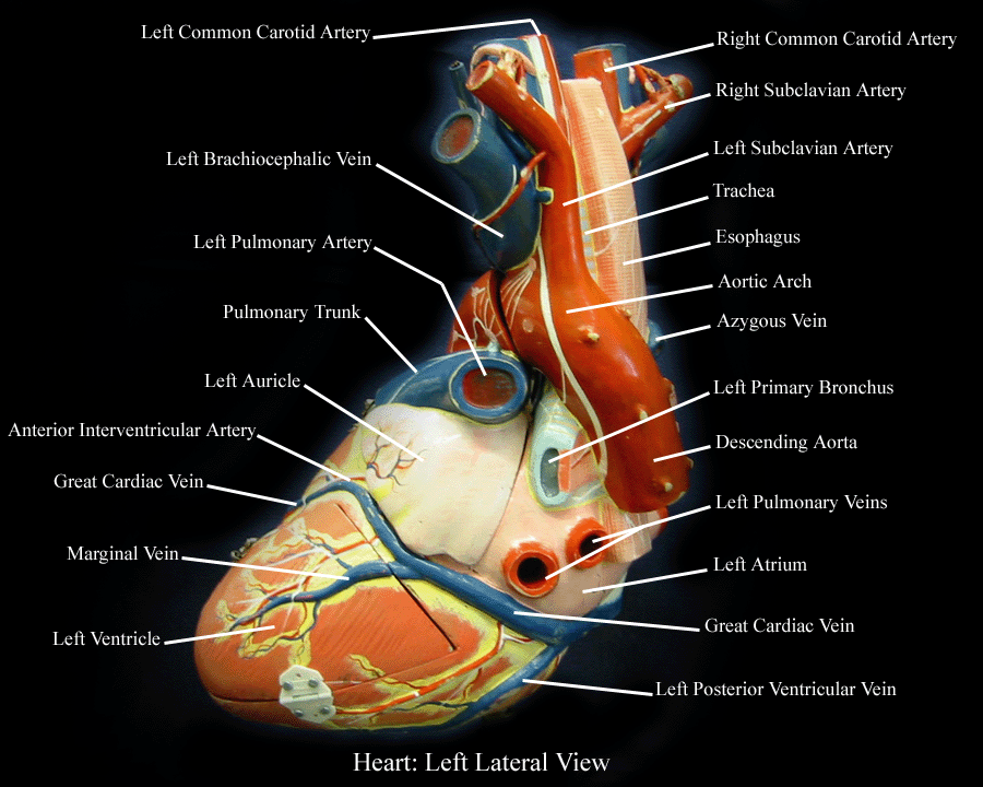
Human Heart Diagram Labeled - Science Trends The heart's atrioventricular valves are structures that join the atria and ventricles of the heart together. This group of valves is comprised of the tricuspid valve and the mitral valve. Beyond this, there is a structure referred to as the aortic valve which separates the left ventricle and the aorta. Cardiovascular System - Heart - Building a Medical Terminology Foundation Specialized groups of heart cells perform this function all on their own, without requiring messages from the central nervous system. Watch this video: Media 12.2. The Heart, Part 2 - Heart Throbs: ... This image shows a magnified view of the structure of the heart wall. Labels read (from top, clockwise): pericardial cavity, fibrous ... Human Heart Diagram Without Labels | Human heart diagram, Heart diagram ... This is Page 32 of a photographic atlas I created as a laboratory study resource for my BIOL 121 Anatomy and Physiology I students on the bones and bony landmarks of the axial skeleton. Credits: All photography, text, and labels by Rob Swatski, Assistant Professor of Biology, Harrisburg Area Community College - York Campus, York, PA. Structure of the Heart | SEER Training The human heart is a four-chambered muscular organ, shaped and sized roughly like a man's closed fist with two-thirds of the mass to the left of midline. The heart is enclosed in a pericardial sac that is lined with the parietal layers of a serous membrane. The visceral layer of the serous membrane forms the epicardium. Layers of the Heart Wall
heart diagram without labels Label The Heart Worksheets (SB6634) - SparkleBox . heart label worksheets diagram human anatomy sparklebox science body ks2 labeling physiology nursing system circulatory diagrams preschool study. Heart anatomy interior labels section cross blood vessels. Free blank heart diagram, download free blank heart diagram png images. Human Heart Diagram Without Labels - Labelling Worksheet This fab labelling worksheet is a great way to help your kids with their learning of the basic human anatomy. It comes with a handy heart diagram without labels, so your students can label it themselves and familiarise themselves with the different parts of the human heart. ear diagram without labels ear diagram without labels Label Parts of the Human Ear we have 9 Images about Label Parts of the Human Ear like Ear Labeling Quiz - Human Anatomy, Label Parts of the Human Ear and also 10 Best Images of Label Ear Diagram Worksheet - Blank Rock Cycle. Here it is: Label Parts Of The Human Ear academic.udayton.edu Human Heart - Diagram and Anatomy of the Heart - Innerbody The heart is a muscular organ about the size of a closed fist that functions as the body's circulatory pump. It takes in deoxygenated blood through the veins and delivers it to the lungs for oxygenation before pumping it into the various arteries (which provide oxygen and nutrients to body tissues by transporting the blood throughout the body).
Natural Science 2: Circulatory system in insects
The Anatomy of the Heart - Quiz 1 - Free Anatomy Quiz The circulatory system - lower body image, with blank labels attached. The circulatory system - a PDF file of the upper and lower body for printing out to use off-line. Describe and explain the function of the circulatory system - The circulatory system consists of the heart, the blood vessels (veins, arteries, and capillaries), and the blood.

Pin on human anatomy organs
Heart: Anatomy and Function - Cleveland Clinic The parts of your heart are like the parts of a house. Your heart has: Walls. Chambers (rooms). Valves (doors). Blood vessels (plumbing). Electrical conduction system (electricity). Heart walls Your heart walls are the muscles that contract (squeeze) and relax to send blood throughout your body.

Ongzi's Lifelong Learning: Typhoid Fever
Diagram of the human heart royalty-free images - Shutterstock 14,700 diagram of the human heart stock photos, vectors, and illustrations are available royalty-free. See diagram of the human heart stock video clips. Set goals and get predicted insights based on performance.

Label Heart Structure | Medical Science Navigator
Heart Blood Flow | Simple Anatomy Diagram, Cardiac Circulation ... - EZmed Step 2 involves the left atrium, the chamber of the heart that receives oxygenated blood from the lungs via the pulmonary veins. 3. Mitral Valve Step 3 involves the mitral valve. During diastole, when the heart is relaxed and filling with blood, the oxygenated blood from the left atrium will flow to the left ventricle.
Heart structure and function - online presentation
Heart Anatomy: Labeled Diagram, Structures, Function, and Blood Flow Chambers of the Heart Let's begin with the chambers of the heart. There are 4 chambers, labeled 1-4 on the diagram below. To help simplify things, we can convert the heart into a square. We will then divide that square into 4 different boxes which will represent the 4 chambers of the heart.
Congenital Heart Defects - How the Heart Works | CDC
Heart Anatomy: size, location, coverings and layers : Anatomy & Physiology Heart Anatomy. The heart is around the size of a fist and weighs between 250-350 grams (less than a pound). Enclosed within the mediastinum, the medial cavity of the thorax, the heart extends obliquely from the second rib to the fifth intercostal space. It rests on the superior surface of the diaphragm, lies posterior to the sternum and ...
32 Label The Diagram Of The Heart - Label Design Ideas 2020
The Heart | Boundless Anatomy and Physiology | | Course Hero Key Points. The heart is a four-chambered muscular organ containing an involuntary conduction system that initiates rhythmic contractions to pump blood throughout the body. The heart has its own blood supply and is controlled by self-regulating nerve bundles called nodes. The SA and AV nodes send impulses through the Purkinje fibers that cause ...
Free Unlabelled Diagram Of The Heart, Download Free Unlabelled Diagram Of The Heart png images ...
The Anatomy of the Heart, Its Structures, and Functions The heart is the organ that helps supply blood and oxygen to all parts of the body. It is divided by a partition (or septum) into two halves. The halves are, in turn, divided into four chambers. The heart is situated within the chest cavity and surrounded by a fluid-filled sac called the pericardium. This amazing muscle produces electrical ...

The structure of the heart - Structure and function of the heart ... The structure of the heart. If you clench your hand into a fist, this is approximately the same size as your heart. It is located in the middle of the chest and slightly towards the left.

The Heart | S-cool, the revision website
Human Heart (Anatomy): Diagram, Function, Chambers, Location in Body Human Heart (Anatomy): Diagram, Function, Chambers, Location in Body The right atrium receives blood from the veins and pumps it to the right ventricle. The right ventricle receives blood from the...

In this diagram they are showing the function of the heart as they have labels to the ...
PDF HEART - STRUCTURE - BiologyMad HEART - STRUCTURE • 4 sections Left atrium Right atrium Left ventricle Right ventricle • heart ry artery Pulmonary vein EAS the blood from he left hand side has to be pumped all around the body. • 2 lo heart Atrioventricular valves - between the atrium and the ventricles Semi-lunar valves - in the pulmonary artery and the aorta

Free Heart Diagram Unlabeled, Download Free Heart Diagram Unlabeled png images, Free ClipArts on ...
the ear diagram without labels ear anatomy. Transparent Digestive System Without Labels ~ News Word lovewordssss.blogspot.com. 60a silica marijuana. 10 Best Images Of Label Ear Diagram Worksheet - Blank Rock Cycle . diagram frog dissection labeled internal label organs toad worksheet heart frogs anatomy structure skeleton labels brain symbol shaped why ear
heart - a level biology student
Heart Diagram with Labels and Detailed Explanation - BYJUS Diagram of Heart. The human heart is the most crucial organ of the human body. It pumps blood from the heart to different parts of the body and back to the heart. The most common heart attack symptoms or warning signs are chest pain, breathlessness, nausea, sweating etc. The diagram of heart is beneficial for Class 10 and 12 and is frequently ...
12+ Model Heart Labeled | Robhosking Diagram
heart without label Vector Winner Golden Label 458912 Vector Art At Vecteezy . winner vector golden label vecteezy. Netizens Have Said TXT YeonJun Looks Like This Former Wanna One Member . txt yeonjun member wanna netizens former said looks kpopmap enregistrée depuis. Normal Chest Axial Anatomy - Plain And Labeled Sections - YouTube

Labeling the Heart
35 Label The Diagram Of The Heart - Labels Design Ideas 2020
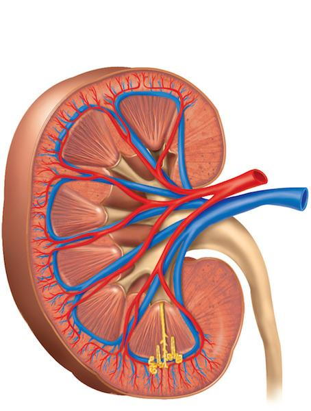
Kidney Disease Facts and Prevention
Post a Comment for "38 structure of the heart without labels"